- Accueil
- Échographie
- Chirurgie mini-invasive par échographie
- Neurological Heel Pain and the Tibial Nerve
Neurological Heel Pain and the Tibial Nerve
Compression of the tibial nerve in the tarsal tunnel of the ankle can lead to heel pain characterized by burning sensations, electric shocks, and sometimes feelings of tingling or numbness. These neuropathic pains, akin to sciatica or cervicobrachial neuralgia, are usually due to the nerve being pinched between the flexor retinaculum of the ankle and the surrounding bony structures.
Dr. Lionel Benamran performs an ultrasound of the tibial nerve to identify potential causes of this compression, such as cysts, supernumerary muscles, or osteoarthritis. An electroneuromyogram can confirm the nerve origin of the pains. While this test can sometimes be negative despite real nerve suffering, an MRI can be helpful in complex cases.
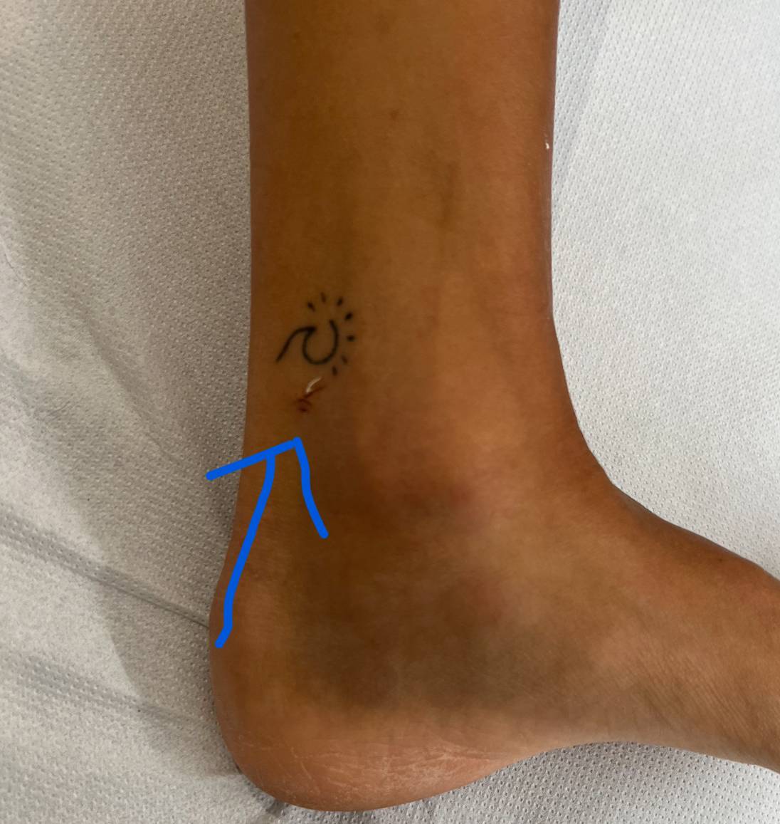
Tibial Nerve Release Surgery in Consultation
Through a minimal incision of 2mm, the surgical release of the tibial nerve is now performed in consultation settings. This procedure, guided by ultrasound, is conducted under local anesthesia using a surgical hook.
This intervention requires no immobilization, and post-surgical pain is typically minimal. Patients can often walk home and return to their work the following day. Local pain due to a hematoma may persist for a few days. The retinaculum heals without recreating pressure but may remain sensitive for about three months.
It is noteworthy that this condition is often associated with plantar fasciitis, whose pathological thickening can also contribute to nerve compression. This can be treated during the same surgical procedure, leaving no visible scar.

 Prendre rendez-vous